
Early Warning Signs Your Tooth Might Need a Root Canal
March 5, 2026Pain that begins inside a tooth often involves the pulp chamber. This inner space contains nerve tissue and blood vessels that support tooth vitality. Bacteria can reach the pulp through untreated decay, cracks, or leaking restorations. Once bacteria enter, inflammation develops inside the confined chamber. Swelling increases pressure against the nerve and may irritate the ligament and bone surrounding the root tip.
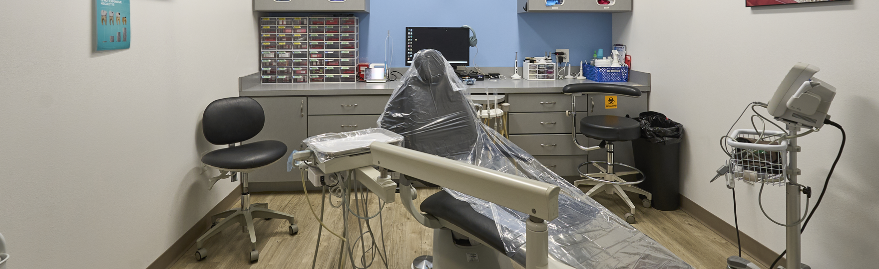
Many people begin searching for root canal treatment near me after discomfort continues for several days or temperature sensitivity becomes prolonged. During consultation, the dentist studies pain patterns, checks gum tissue around the tooth, and reviews dental radiographs. These observations help determine whether inflammation remains limited to the pulp or whether bacterial infection has reached the bone around the root.

Persistent Tooth Pain That Doesn’t Go Away on Its Own
Long-lasting tooth pain often indicates irritation within the pulp tissue. Bacterial toxins trigger inflammation inside the chamber, and pressure builds around nerve fibers. Because enamel and dentin cannot expand, the irritated nerve continues sending pain signals.
Several clinical findings guide the dentist’s judgment:
- Discomfort that increases during chewing or biting pressure
- Sensitivity when the tooth is lightly tapped
- Radiographic signs of inflammation near the root tip
These findings help distinguish reversible irritation from irreversible pulp damage. If nerve tissue remains capable of healing, treatment may focus on removing decay and sealing the tooth. When the pulp no longer functions normally, removing infected tissue prevents bacteria from moving deeper into the root canal system.
Sensitivity to Hot or Cold That Lingers After Eating or Drinking
Temperature sensitivity can occur when dentin transmits thermal changes toward the nerve. In healthy teeth, the sensation fades quickly once the stimulus disappears.
Inflamed pulp behaves differently. Irritated nerve fibers recover more slowly, allowing the sensation to persist even after the hot or cold source is removed. Dentists near me observe how long the response lasts during controlled temperature testing.
Important observations include:
- Duration of the sensation after the stimulus ends
- Changes in discomfort during repeated testing
- Signs of inflammation in tissues near the root tip
Prolonged sensitivity often signals that the pulp cannot recover without intervention. When inflammation continues, removing infected tissue may protect the surrounding bone and ligament structures.
Swelling, Tender Gums, or Pimple-Like Bumps Near the Tooth
In some cases, bacteria travel through the root canal and reach the bone at the end of the root. The immune system reacts by producing inflammatory fluid. Pressure from this fluid may create localized swelling in nearby gum tissue.
Occasionally, a small drainage pathway forms through the gum surface. This opening may resemble a pimple or small bump near the affected tooth. Fluid may escape through the opening, but bacteria can remain inside the root canal space.
During examination, the dentist looks for:
- Local swelling near the root area
- Gum tenderness when light pressure is applied
- Drainage points that suggest infection in bone tissue
Radiographs help reveal whether the bone around the root tip has begun to change. Treatment planning depends on how far the infection has progressed and whether the tooth remains stable in the surrounding bone. If swelling or drainage near a tooth continues, it may be appropriate to reach out to a dentist in Princeton, TX, for a clinical examination and imaging to determine whether the infection involves the root canal system.
Tooth Discoloration and What It May Indicate Internally
A tooth that gradually darkens may indicate damage within the pulp chamber. Trauma, deep cavities, or prolonged inflammation can disrupt the small blood vessels inside the pulp.
Breakdown products from injured pulp tissue may diffuse into dentin and alter the color of the tooth. As this process continues, the affected tooth may appear gray, brown, or darker than neighboring teeth.
Clinical evaluation focuses on several indicators:
- Nerve response during vitality testing
- Radiographic evidence of changes near the root tip
- Condition of the ligament supporting the tooth
Discoloration alone does not confirm infection. However, a color change combined with pain or lingering sensitivity raises concern about internal pulp damage.
How Dentists Diagnose Infection Inside the Tooth?
Diagnosis requires several clinical procedures that reveal the condition of nerve tissue and surrounding structures. Each test contributes information that guides treatment decisions.
Typical diagnostic steps include:
- Visual inspection to identify decay, fractures, or leaking restorations
- Thermal testing to observe nerve response to temperature changes
- Percussion testing to detect inflammation in the periodontal ligament
- Dental radiographs to identify infection near the root tip or changes in bone density
Dentists interpret these findings together rather than relying on a single test. Limited nerve response combined with bone changes near the root often indicates that the pulp is no longer vital.
Why Early Root Canal Treatment Can Save Your Natural Tooth?
Once bacteria infect the pulp, the root canal system becomes a pathway for microorganisms to reach the surrounding bone. Root canal therapy focuses on removing infected tissue and disinfecting the internal canal space while preserving the tooth root.
The procedure generally includes several controlled steps:
- Creating an opening into the pulp chamber
- Removing damaged nerve tissue
- Cleaning and shaping the internal canal system
- Disinfecting and sealing the canals
After sealing the canal system, the dentist checks the remaining tooth structure. Teeth weakened by decay or internal damage may require a crown to stabilize chewing forces. Individuals who ask about root canal treatment services are first examined to determine whether the root and surrounding bone can support long-term healing.
Experiencing These Symptoms? Schedule a Dental Evaluation Before the Problem Worsens
Persistent pain, prolonged sensitivity, or swelling near a tooth may indicate inflammation within the pulp. Dentists review nerve vitality, examine gum tissue, and analyze dental imaging to understand the condition of the root and surrounding bone.
Early clinical evaluation clarifies whether the pulp may recover or whether infection requires treatment. Individuals seeking care from a dentist may undergo diagnostic examination at Hello Dental & Orthodontics, where the condition of the tooth, ligament, and bone support is carefully reviewed before treatment decisions are made.