
Why Regular Dental Exams Detect More Than Cavities?
April 25, 2026A dental examination involves more than identifying cavities. Teeth are supported by bone, connective fibers, and gum tissue. Bacteria accumulating along the gum line can irritate these structures and lead to inflammation that slowly weakens bone around the roots.
Many adults begin searching online for a tooth exam near me only after swelling, bleeding, or sensitivity develops. At that point, decay or periodontal infection may already involve deeper layers such as dentin or surrounding bone. Regular dental exams allow the clinician to inspect enamel surfaces, measure gum attachment, review bone height on radiographs, and observe how the upper and lower teeth contact during chewing. Findings from these observations guide decisions about preventive care or treatment.

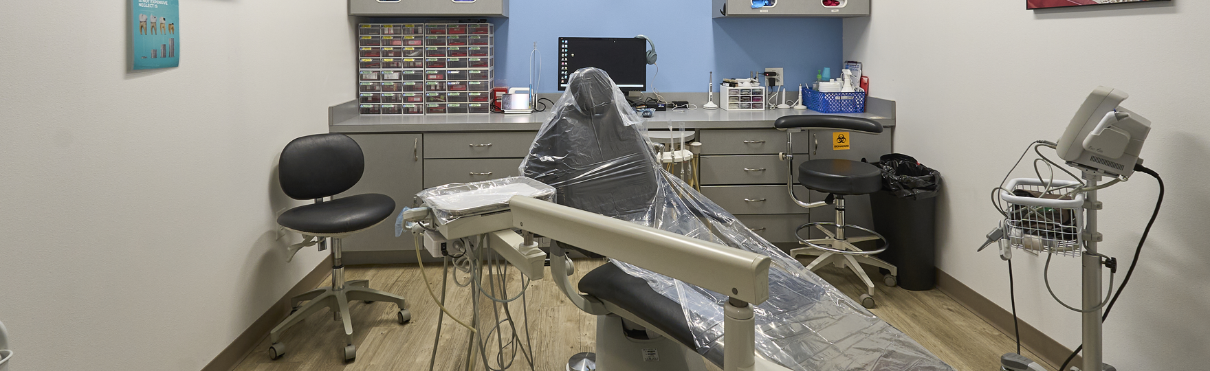
What Happens During a Comprehensive Dental Examination?
A full dental exam reviews several structures inside the mouth because tooth stability depends on the surrounding bone and soft tissue. Each step focuses on identifying structural damage, bacterial buildup, or changes in alignment.
Typical examination steps include:
- Inspection of tooth surfaces to identify softened enamel, fractures, or early decay that may expose dentin.
- Periodontal probing, where a small measuring instrument checks the depth between the tooth and the gum tissue to assess the attachment of connective fibers.
- Dental radiographs are used to review bone height around roots and identify infection that may form near the root tip.
- Soft-tissue inspection of the tongue, cheeks, palate, and throat to identify abnormal patches or persistent irritation.
- Bite analysis to observe whether chewing forces distribute evenly across the dental arch.
During routine dental checkup services, these observations are recorded and compared with previous records. Changes in bone levels, gum attachment, or enamel wear may suggest bacterial activity, excessive bite pressure, or structural weakness in a tooth.
How Routine Exams Help Identify Gum Disease Early?
Gum disease begins with plaque accumulation along the margin where the tooth meets the gum. The immune response to bacteria produces redness and swelling in the gingival tissue. If inflammation persists, connective fibers that anchor the tooth to the bone may weaken.
Clinical review focuses on several measurable findings:
- Depth of the pocket between the gum and the tooth root
- Bleeding during the gentle probing of the gum tissue
- Areas where the gum has receded and exposed root surfaces
- Bone levels around the teeth on radiographic images
If inflammation appears limited to the gum surface, removal of plaque and hardened tartar may allow the tissue to recover. When bacteria extend deeper beneath the gum margin, scaling and root cleaning may be recommended. Bone support and pocket depth are checked again during future visits to observe whether healing has occurred.
Detecting Oral Cancer and Other Hidden Health Concerns
Soft tissue inside the mouth can show changes that are not immediately painful. Dentists inspect the tongue, cheeks, lips, and floor of the mouth because abnormal tissue growth may begin in these areas.
During this screening, the clinician checks for:
- Red or white patches that remain after normal healing
- Firm lumps beneath the mucosal surface
- Ulcers that persist longer than expected
- Thickened or rough areas of tissue
Some findings represent irritation from biting trauma or infection. Other lesions may require closer observation. If tissue changes remain during follow-up visits, additional evaluation or referral for biopsy may be considered depending on the location and appearance of the area.
Signs of Teeth Grinding, Bite Issues, and Jaw Problems
Alignment of the teeth influences how chewing pressure spreads across enamel and bone. Repeated grinding places heavy mechanical force on tooth surfaces and jaw joints.
Dentists look for structural clues such as:
- Flattened chewing surfaces from grinding pressure
- Fine fractures in enamel extending toward dentin
- Tender jaw muscles near the temporomandibular joint
- Uneven bite contact that concentrates force on certain teeth
These findings help determine whether chewing forces are balanced. Excessive pressure on a limited number of teeth may lead to cracks, sensitivity, or joint discomfort. Protective night appliances may be considered in some cases to reduce stress on enamel and stabilize the bite.
How Dental Exams Reveal Problems Before Pain Starts?
Pain often starts after bacteria spread into the pulp, the inner tissue of the tooth where nerves and blood vessels are located. Earlier stages of decay may affect enamel and dentin without producing noticeable symptoms.
Careful inspection and radiographic imaging allow dentists to identify these changes earlier. Small cavities sometimes appear on X-ray images before visible damage develops on the surface. Hairline enamel cracks may also become visible under strong lighting or magnification during examination.
Recognizing these structural changes early allows the dentist to decide whether the tooth requires monitoring, remineralization therapy, or removal of decayed material before infection approaches the nerve.
How Oral Health Relates to Overall Health
Gum tissue contains small blood vessels that connect the mouth with the rest of the body. When the gums are inflamed, oral bacteria may enter these vessels during chewing or brushing. For this reason, periodontal infection is reviewed carefully during routine dental visits.
Bone loss around teeth usually reflects long-term inflammation affecting the surrounding tissue. Examination of gum attachment, plaque levels, and tissue healing helps determine whether bacterial activity remains active or has improved following cleaning and daily oral care.
Dental professionals often coordinate oral findings with a patient’s general medical history when inflammation persists or healing appears delayed.
Stay Ahead of Dental Problems — Schedule Your Regular Dental Exam Today
Regular dental exams allow changes in tooth structure, gum attachment, and bone support to be observed over time. Subtle differences in enamel wear, pocket depth, or bite pressure may signal early disease or mechanical stress affecting the teeth.
Adults searching for a dentist in Princeton, TX often schedule periodic examinations so these structures can be reviewed before symptoms develop. Clinical observations collected during these visits guide decisions about cleaning, repair, or continued observation depending on bone stability and bacterial activity.
Comprehensive oral evaluations performed at Hello Dental & Orthodontics focus on inspecting tooth structure, measuring gum attachment, reviewing bone levels, and identifying infection before it progresses toward nerve involvement or structural tooth damage.