
What to Expect During Your First Cosmetic Dentistry Consultation in Princeton, TX
April 10, 2026A cosmetic consultation begins with a careful review of tooth structure, gum tissue, and the bone that supports each root. Many adults schedule this visit after searching for cosmetic dentistry near me because they notice staining, uneven edges, or changes in alignment. Cosmetic changes can only remain stable when the surrounding tissues are healthy.
Each tooth sits in bone and connects to the surrounding tissue through periodontal ligaments and nerve pathways. If inflammation or infection affects these structures, restorative work may fail, or healing may slow. For that reason, the dentist studies the enamel condition, gum attachment, and bite pressure before discussing cosmetic procedures. Findings from this evaluation guide the clinical decision about whether cosmetic treatment can proceed or whether preventive care must stabilize the tissues first.

Understanding Your Smile Goals and Cosmetic Concerns
Early discussion focuses on identifying the concern and linking it to a biological cause. Changes in color, spacing, or shape may develop from enamel erosion, minor fractures, shifting teeth, or gum recession that exposes root surfaces.
Visible cosmetic issues often connect to underlying structural changes in the tooth or surrounding tissue. Enamel loss may uncover dentin, which contains microscopic tubules connected to nerves inside the tooth. This exposure can produce sensitivity when pressure or temperature changes occur.
During this stage, the dentist reviews several clinical observations:
- enamel wear that may expose dentin and increase nerve response
- gum recession that reduces tissue protection around the root
- crowding or spacing that traps plaque and increases infection risk
- uneven bite contact that concentrates chewing pressure on certain teeth
These findings help determine whether cosmetic dental services address the concern directly or whether additional treatment should occur before cosmetic work begins.
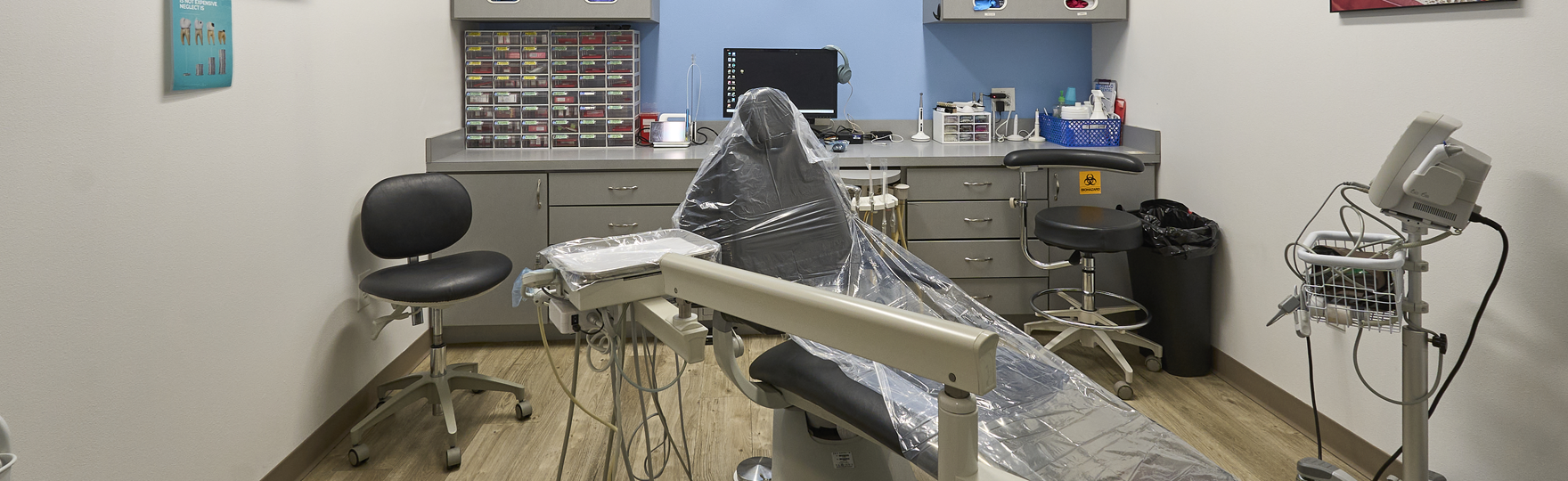
Comprehensive Oral Examination and Smile Evaluation Process
A structured oral examination follows the discussion. Each tooth is inspected along with the surrounding gum tissue and bone support that anchors the root. Strong bone support allows teeth to withstand normal chewing pressure after cosmetic restoration.
Radiographs may be taken when the root structure or bone level requires closer inspection. These images reveal bone height around the root and help identify decay or infection that may exist beneath the gum line.
Several clinical checkpoints guide the examination:
- gum tissue condition, including swelling or bleeding
- enamel integrity, including fractures or areas of decay
- bite contact between upper and lower teeth
- condition of existing fillings, crowns, or bonding materials
If periodontal infection or bone loss appears during this examination, the dentist will address those conditions first. Cosmetic procedures rely on stable gum tissue and adequate bone support to remain functional.
Digital Imaging and Smile Design Preview Options
Digital photographs and intraoral scans provide detailed records of tooth position, gum contour, and alignment within the dental arch. These records allow the dentist to examine the relationship between teeth, soft tissue, and underlying bone.
Image analysis helps determine whether the enamel layer allows minor reshaping or veneer preparation. Removing too much enamel can expose dentin and weaken the tooth structure, so careful inspection of tooth thickness becomes important.
Computer simulations may demonstrate how changes in tooth length, contour, or alignment could affect the overall smile. These images serve as visual references rather than predictions of final results. The dentist studies these previews while considering biological limits such as gum position, bite pressure, and bone support.
Digital records also allow comparison during later visits to confirm that restorations fit properly and that surrounding tissues remain stable.
Exploring Cosmetic Treatment Options Based on Your Needs
Treatment recommendations depend on enamel strength, gum stability, and the way chewing forces distribute across the bite. Cosmetic procedures either modify the outer enamel surface or place restorative material that bonds to the tooth.
Possible approaches may include:
- teeth whitening when discoloration affects enamel without structural damage
- dental bonding for small fractures or minor gaps between teeth
- porcelain veneers are used when the enamel thickness supports conservative preparation
- clear aligners, when mild tooth movement improves alignment and bite balance
Each procedure must function within biological limits. Teeth that receive excessive bite pressure may require alignment correction before cosmetic restoration. Gum recession or enamel thinning may also influence which procedures remain appropriate. A cosmetic dental clinic reviews these structural factors because cosmetic materials must tolerate everyday chewing forces without damaging the tooth or surrounding tissue.
Discussing Treatment Timeline, Costs, and Financing Options
The sequence of treatment depends on biological healing and the condition of the surrounding tissues. Certain cosmetic procedures can occur during a single appointment, while others require several visits so gum tissue and bite alignment can stabilize between steps.
Dentists near me typically explain several practical details:
- approximate duration of each procedure
- situations where temporary restorations protect the tooth during healing
- follow-up visits are needed to observe the gum tissue response
- financial planning when treatment involves multiple stages
If inflammation or infection appears during the examination, cosmetic procedures may be postponed until the tissues recover. Decisions about timing depend on how the gums, bone, and tooth structure respond to earlier treatment.
Creating a Personalized Smile Makeover Plan
After completing the examination and reviewing treatment options, the dentist organizes a treatment sequence that protects tooth structure and supporting tissues. Alignment, gum stability, and bone support must remain consistent before cosmetic restorations are placed.
A treatment plan may involve several coordinated steps:
- stabilizing gum tissue and controlling infection if present
- correcting alignment when bite pressure concentrates on certain teeth
- restoring enamel shape through bonding or ceramic restorations
- monitoring tissue response and bite balance after placement
Follow-up visits allow the dentist to observe how restorations interact with chewing pressure. Adjustments may occur if the bite shifts or if gum tissue changes position during healing.
Ready to Enhance Your Smile? Schedule Your Cosmetic Dentistry Consultation in Princeton, TX Today
Cosmetic treatment begins with evaluation of tooth structure, gum attachment, and bone support. A dentist in Princeton, TX, reviews these biological factors before recommending cosmetic procedures. Bone level around the roots, tissue health, and bite pressure influence whether cosmetic restorations remain stable over time.
Clinical consultations at Hello Dental & Orthodontics involve a systematic examination of enamel condition, periodontal tissue, and alignment before any cosmetic procedure is considered. Decisions depend on structural stability, healing capacity, and the interaction between teeth and surrounding tissue.