
What Your Dentist Looks for During Oral Cancer Screenings?
April 15, 2026Routine dental examinations include inspection of the soft tissues lining the mouth because abnormal cellular activity can interrupt normal healing and sometimes extend into nearby bone or lymphatic structures. Some people search for an oral cancer screening near me, although this type of evaluation is often included during standard dental visits. During the appointment, the dentist examines the cheeks, tongue, palate, and throat to assess tissue color, thickness, and surface integrity. Healthy mucosal tissue should appear uniform and flexible. Areas that show persistent redness, white patches, or delayed healing require observation because these changes can indicate irritation, infection, or abnormal cell development affecting the surrounding tissue.
Why Oral Cancer Screenings Are an Important Part of Routine Dental Visits?
Oral tissues regenerate through a continuous cycle in which epithelial cells replace older surface cells. Stable regeneration depends on proper blood circulation and intact connective tissue beneath the mucosal surface. Disruption in this process can lead to lesions that do not heal normally. Because early changes may develop without pain or nerve involvement, routine dental visits provide an opportunity to examine the tissue before symptoms appear.
During the clinical examination, the dentist studies the surface of the oral mucosa and assesses whether the tissue maintains normal elasticity and coloration. Ulcers that remain longer than expected, areas of thickened mucosa, or unexplained discoloration may signal irritation or cellular change. When such findings appear, consulting a dentist near me can help ensure that appropriate monitoring or further diagnostic evaluation is considered based on the duration and characteristics of the lesion.
Common Warning Signs Dentists Check Inside the Mouth and Throat
A careful oral examination includes visual inspection combined with gentle palpation of soft tissues. Changes within epithelial layers or underlying connective structures can affect normal healing and may require closer observation.
Dentists commonly assess the following signs:
- Red or white patches that indicate altered mucosal tissue
- Ulcers that persist longer than expected or fail to heal
- Thickened areas that change the normal texture of the cheek lining or tongue
- Swelling that interferes with jaw movement or soft tissue stability
- Lumps in the neck that may involve lymph nodes
Each finding is evaluated based on location, size, and duration. If the tissue does not return to a normal appearance after a short observation period, additional testing or referral for biopsy may be considered to determine the cause of the change.
Factors That May Increase the Risk of Oral Cancer
Oral tissues respond to repeated irritation, infection, and environmental exposure. Dentists review health history because certain conditions can weaken the protective mucosal lining and influence how tissue heals after injury.
Several factors are known to affect tissue health:
- Tobacco exposure can damage epithelial cells and delay healing
- Alcohol consumption, which may irritate oral tissues and reduce protective barriers
- Human papillomavirus (HPV) infection, which can influence cellular growth in the throat tissue
- Long-term sun exposure that affects the lips
- Reduced immune function that may limit the body’s response to infection
These factors do not confirm the disease. Instead, they guide the dentist in determining whether oral tissues require closer observation or more frequent examination.
How Early Detection Improves Treatment Outcomes?
Cellular abnormalities in the mouth often begin in the outer epithelial layers. At this stage, the affected area may remain localized within a small section of mucosa. Identifying the change early allows healthcare providers to assess the lesion before deeper tissue structures become involved.
Progression of abnormal cells may extend into connective tissue, bone, or nearby lymphatic pathways. Once deeper structures become affected, treatment decisions can become more complex because a larger area of tissue may require removal or stabilization. Early identification allows clinical teams to evaluate the lesion while surrounding bone, nerves, and functional structures remain intact.
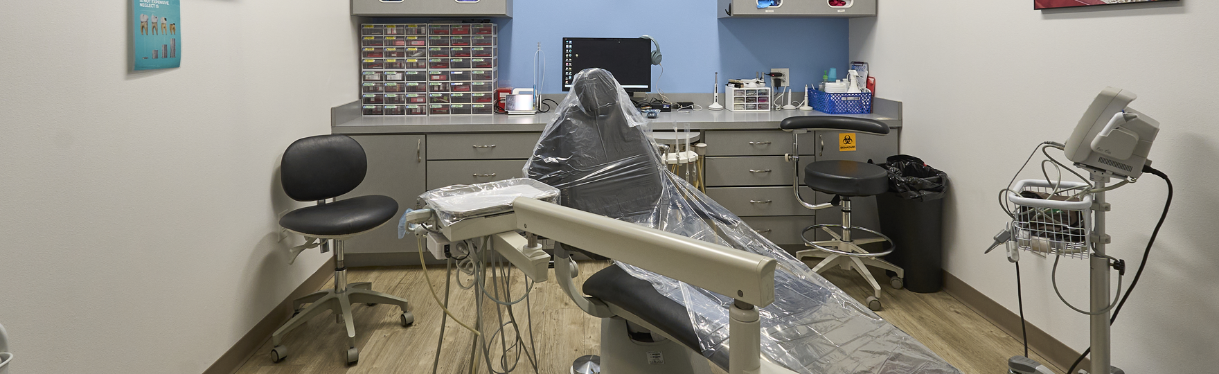
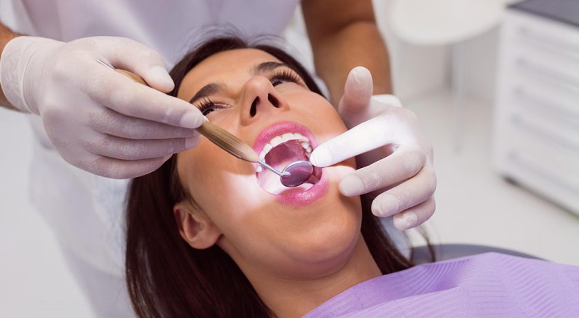
What Happens During a Professional Oral Cancer Screening Exam?
A clinical screening focuses on systematic inspection of the oral cavity and surrounding structures. The dentist examines visible tissue surfaces and palpates underlying areas to detect abnormalities that may not be immediately visible.
The assessment usually includes:
- Inspection of the lips, cheeks, tongue, and palate to observe color and surface texture
- Palpation of the floor of the mouth to detect thickened tissue or small nodules
- Examination of the throat and tonsil region for irregular swelling
- Evaluation of the neck to determine whether lymph nodes appear enlarged
Some dental practices include visualization devices during oral cancer screening services. These lights can reveal differences in tissue fluorescence, which may help identify areas that require closer examination. If irregular tissue appears, the dentist evaluates whether monitoring is appropriate or whether referral for biopsy should be considered based on the appearance and duration of the lesion.
How Frequently Should Oral Cancer Screenings Be Performed?
Oral tissue changes often develop gradually. Periodic monitoring helps identify differences that might not be noticeable during a single examination. Many adults receive screening during routine dental visits approximately every six months. This interval allows comparison of tissue appearance and healing patterns between appointments.
Certain health factors may require closer observation. Tobacco exposure, previous abnormal lesions, or viral infections may influence the frequency of tissue evaluation. The dentist reviews these factors and determines whether additional follow-up visits are appropriate, depending on how the oral tissues respond over time.
Protect Your Health with Early Detection — Schedule Your Oral Cancer Screening Today
Regular evaluation of oral tissues helps identify structural changes that may interfere with healing or affect nearby bone and nerve structures. During these examinations, the dentist examines tissue color, thickness, and surface texture to determine whether abnormalities require additional investigation. If an unusual lesion appears, the area may be monitored for healing or referred for further testing, depending on clinical findings. Individuals seeking care from a dentist in Princeton, TX often receive this evaluation during routine examinations, and clinical visits at Hello Dental & Orthodontics include a systematic assessment of oral tissues so any irregular cellular changes can be identified and reviewed appropriately.